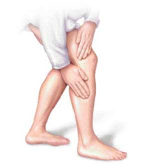
Beside cancer, heart disease kills more than 2,000 Americans everyday. Approximately 60 million Americans have heart disease.I. Causes of Heart DiseasesThere are many causes of heart diseases. Most of heart diseases are caused by high blood pressure contributes to hardening of the arteries. High levels of bad cholesterol (LDL) build up in the arteries as a result of uncontrolled diet with high levels of saturated fat and trans fat. All these add to the formation of atherosclerosis lesions and eventually arterial blockage or anything that serves to damage the inner lining of blood vessels and impedes the transportation of oxygen and nutrition to the heart can be defined as a risk of heart disease.II Symptoms of Heart diseasesBeside cancer, heart disease kills more than 2,000 Americans everyday. Approximately 60 million Americans have heart disease. There are many causes of heart disease. Anything that serves to damage the inner lining of blood vessels and impedes the transportation of oxygen and nutrition to the heart can be defined as a risk of heart disease. Here are some early indication of heart disease symptoms:1. Leg cramps during walkingLeg cramps during exercise might be caused by dehydration. It is important to drink a lot of fluid during exercise. Leg cramps occur when the muscle suddenly and forcefully contracts. The most common muscles to contract in this manner are muscles that cross two joints. Leg cramps during walking might be an indication of heart disease caused by arteries in your leg being clogged up by cholesterol in result of not enough oxygen being delivered to the cells in your leg. If this symptom persists, please consult with your doctor.2. Chest painChest pain is caused by blood vessels in the heart temporarily being blocked up. It is also caused by inadequate oxygen supply to the heart muscle or coronary . The persistence of chest pain would be an early indication of heart diseases.3. Shortness of breathShortness of breath (dyspnea) is the major symptom of the left ventricular insufficiency. People with shortness of breath are four times more likely to die from a heart disease related cause than individuals without any symptoms.4. HeadachesPeople see sparkling zigzag lines or loss of vision before a migraine attack may be at particular risk of future cardiovascular problems. Generally headaches do not cause heart diseases but a sudden, explosive onset of great pain might be.5. DizzinessDizziness can have many causes including low blood count, low iron in the blood stream and other blood disorders, dehydration, and viral illnesses. Since there are many different conditions that can produce these symptoms, anybody experiencing episodes of severe headaches or dizziness ought to be checked by your doctor.6. PalpitationsPalpitations is an extremely common symptom of heart disease. Palpitations are skips in the heart beats and irregular heart beats.7. Loss of consciousnessIt is a common symptom, most people pass out at least once in their lives. However, sometimes loss of consciousness indicates a dangerous or even life-threatening condition such as heart disease so when loss of consciousness occurs it is important to figure out the cause.There are many more symptoms such as fatigue, memory defects, and changes in skin tone and temperature.III. Types of Heart DiseasesThe heart is a four chambered, hollow muscle and double acting pump that is located in the chest between the lungs. Heart diseases caused by high blood pressure contributes to hardening of the arteries. High levels of bad cholesterol (LDL) build up in the arteries as a result of uncontrolled diet with high levels of saturated fat and trans fat. All these add to the formation of atherosclerosis lesions and eventually arterial blockage.There are some major types of heart diseases:1. Type of heart disease affecting heart chambersAs we mention in the previous article, the heart is a four chambered hollow muscle and double acting pump that is located in the chest between the lungs. Heart diseases caused by high blood pressure contributes to hardening of the arteries. High levels of bad cholesterol (LDL) build up in the arteries as a result of uncontrolled diet with high levels of saturated fat and trans fat. All these add to the formation of atherosclerosis lesions and eventually arterial blockage.In this article, we will discuss heart disease affecting the heart chambers.Heart failure is caused by the heart not pumping as much blood as it should and so the body does not get as much blood and oxygen that it needs. The malfunctioning of the heart chambers are due to damage caused by narrowed or blocked arteries leading to the muscle of your heart.There are 4 heart chambers as follow: * The right atrium * The left atrium * The right ventricle * The left ventricle.Heart diseases affect the heart chambers include:A. Congestive heart failureHeart failure is caused by the heart not pumping as much blood as it should and so the body does not get as much blood and oxygen that it needs. The malfunctioning of the heart chambers are due to damage caused by narrowed or blocked arteries leading to the muscle of your heart.a) Diastolic dysfunction:The contraction function is normal but there's impaired relaxation of the heart, impairing its ability to fill with blood causing the blood returning to the heart to accumulate in the lungs or veins.b) Systolic dysfunction:The relaxing function is normal but there's impaired contraction of the heart causing the heart to not pump out as much blood that is returned to it as it normally does as a result of more blood remaining in the lower chambers of the heart.B. Pulmonary heart diseasePulmonary heart disease is caused by an enlarged right ventricle. It is known as heart disease resulting from a lung disorder where the blood flowing into the lungs is slowed or blocked causing increased lung pressure. The right side of the heart has to pump harder to push against the increased pressure and this can lead to enlargement of the right ventricle.2. Heart Disease affecting heart musclesIn the case of heart diseases affecting heart muscles, the heart muscles are stiff, increasing the amount of pressure required to expand for blood to flow into the heart or the narrowing of the passage as a result of obstructing blood flow out of the heart.Heart diseases affecting heart muscles include:1. CardiomyopathyHeart muscle becomes inflamed and doesn't work as well as it should. There may be multiple causes such as high blood pressure, heart valve disease, artery diseases or congenital heart defects.a) Dilated cardiomyopathyThe heart cavity is enlarged and stretched. Blood flows more slowly through an enlarged heart, causing formation of blood clots as a result of clots sticking to the inner lining of the heart, breaking off the right ventricle into the pulmonary circulation in the lung or being dislodged and carried into the body's circulation to form emboli .b) Hypertrophic cardiomyopathyThe wall between the two ventricles becomes enlarged, obstructing the blood flow from the left ventricle. Sometimes the thickened wall distorts one leaflet of the mitral valve, causing it to leak. The symptoms of hypertrophic cardiomyopathy include shortness of breath, dizziness, fainting and angina pectoris.c) Restrictive cardiomyopathyThe ventricles becomes excessively rigid, so it's harder for the ventricles to fill with blood between heartbeats. The symptoms of restrictive cardiomyopathy include shortness of breath, swollen hands and feet.2. Myocarditis Myocarditis is an inflammation of the heart muscles or the weaken of the heart muscles. The symptoms of myocarditis include fever, chest pains, congestive heart failure and palpitation.3. Heart disease affecting heart valvesHeart diseases affecting heart valves occur when the mitral valve in the heart narrows, causing the heart to work harder to pump blood from the left atrium into the ventricle.Here are some types of heart disease affecting heart valves:1. Mitral StenosisMitral Stenosis is a heart valve disorder that involves a narrowing or blockage of the opening of the mitral valve causing the volume and pressure of blood in the left atrium increases.2. Mitral valves regurgitationMitral regurgitation is the heart disease in which your heart's mitral valve doesn't close tightly causing the blood to be unable to move through the heart efficiently. Symptoms of mitral valve regurgitation are fatigue and shortness of breath.3. Mitral valves prolapseIn mitral valve prolapse, one or both leaflets of the valve are too large resulting in uneven closure of the valve during each heartbeat. Symptoms of mitral valves prolapse are palpitation, shortness of breath, dizzy, fatigue and chest pains.4. Aortic StenosisWith aging, protein collagen of the valve leaflets are destroyed and calcium is deposited on the leaflets causing scarring, thickening, and stenosis of the valve therefore increasing the wear and tear on the valve leaflets resulting in the symptoms and heart problems of aortic stenosis.5. Aortic regurgitationAortic regurgitation is the leaking of the aortic valve of the heart that causes blood to flow in the reverse direction during ventricular diastole, from the aorta into the left ventricle. Symptoms of aortic regurgitation include fatigue or weakness, shortness of breath, chest pain, palpitation and irregular heart beats.6. Tricuspid stenosisTricuspid stenosis is the narrowing of the orifice of the tricuspid valve of the heart causing increased resistance to blood flow through the valve. Symptoms of tricuspid stenosis include fatigue, enlarged liver, abdominal swelling, neck discomfort and leg and ankle swelling.7. Tricuspid regurgitation.Tricuspid regurgitation is the failure of the right ventricular causing blood to leak back through the tricuspid valve from the right ventricle into the right atrium of the heart. Symptoms of tricuspid regurgitation include leg and ankle swelling and swelling in the abdomen.4. Heart disease affecting coronary arteries and coronary veinsHeart disease affecting coronary arteries and coronary veins:The malfunctioning of the heart may be due to damage caused by narrowed or blocked arteries leading to the muscle of your heart as well as blood backing up in the veins. Types of heart disease that affect the coronary arteries and veins include:A. Angina pectorisAngina pectoris occurs when the heart muscle doesn't get as much blood oxygen as it needs. Here are 3 types of angina pectoris:a) Stable anginaStable angina is chest pain or discomfort that typically occurs with activity or stress due to oxygen deficiency in the blood muscles and usually follows a predictable pattern. Symptom of stable angina include chest pain, tightness, pressure, indigestion feeling and pain in the upper neck and arm.b) Unstable anginaUnstable angina is caused by blockage of the blood flow to the heart. Without blood and the oxygen, part of the heart starts to die. Symptoms of unstable angina include pain spread down the left shoulder and arm to the back, jaw, neck, or right arm, discomfort of chest and chest pressure.c) Variant angina also known as coronary artery spasmCaused by the narrowing of the coronary arteries. This is caused by the contraction of the smooth muscle tissue in the vessel walls. Symptoms of variant angina include increasing of heart rate, pressure and chest pain.B. Heart attacks known as myocardial infarction or MIHeart attacks caused by plaque rupture with thrombus formation in a coronary vessel, resulting in an acute reduction of blood supply to a portion of the myocardium. Symptoms of MI include a squeezing sensation of the chest, sweating, nausea and vomiting, upper back pain and arm pain.C. Heart disease also known as coronary artery disease or coronary heart diseaseCaused by arteries hardening and narrowing, cutting off blood flow to the heart muscle and resulting in heart attack. Symptoms of heart disease include shortness of breath, chest pains on exertion, palpitation, dizziness and fainting.D. Atherosclerosis or hardening of arteriesArteries are blood vessels that carry oxygen-rich blood to your heart and to other parts of your body. Atherosclerosis is caused by plaques that rupture in result of blood clots that block blood flow or break off and travel to another part of the body. Atherosclerosis has no symptom or warning sign.E. Silent ischemia.Ischemia is a condition in which the blood flow is restricted to a part of the body caused by narrowing of heart arteries. Silent ischemia means people have ischemia without pain. There is also no warning sign before heart attack.5. Heart disease affecting heart liningRheumatic heart disease results from inflammation of the heart lining when too much fluid builds up in the lungs leading to pulmonary congestion. It is due to failure of the heart to remove fluid from the lung circulation resulting in shortness of breath, coughing up blood, pale skin and excessive sweating. Heart disease resulting from inflammation of either the endocardium or pericardium is called heart disease affecting heart lining.Endocardium is the inner layer of the heart. It consists of epithelial tissue and connective tissue. Pericardium is the fluid filled sac that surrounds the heart and the proximal ends of the aorta, vena vava and the pulmonary artery.1. EndocarditisEndocarditis, which is an inflammation of the endocardium is caused by bacteria entering the bloodstream and settling on the inside of the heart, usually on the heart valves that consists of epithelial tissue and connective tissue. It is the most common heart disease in people who have a damaged, diseased, or artificial heart valve. Symptoms of endocarditis include fever, chilling, fatigue, aching joint muscles, night sweats, shortness of breath, change in temperature and a persistent cough.2. PericardiumPericarditis is the inflammation of the pericardium. It is caused by infection of the pericardium which is the thin, tough bag-like membrane surrounding the heart. The pericardium also prevents the heart from over expanding when blood volume increases. Symptoms of pericarditis include chest pain, mild fever, weakness, fatigue, coughing, hiccups, and muscle aches.6. Heart disease affecting electrical systemThe electrical system within the heart is responsible for ensuring the heart beats correctly so that blood can be transported to the cells throughout our body. Any malfunction of the electrical system in the heart causes a fast, slow, or irregular heartbeat. The electrical system within the heart is responsible for ensuring that the heart beats correctly so that blood can be transported throughout our the body. Any malfunction of the electrical system in the heart malfunction can cause a fast, slow, or irregular heartbeat.Types of heart disease that affect the electrical system are known as arrhythmias. They can cause the heart to beat too fast, too slow, or irregularly. These types of heart disease include:a. Sinus tachycardiaSinus tachycardia occurs when the sinus rhythm is faster than 100 beats per minute therefore it increases myocardial oxygen demand and reduces coronary blood flow, thus precipitating an ischemia heart or valvular disease.b. Sinus bradycardiaSinus bradycardia occurs when a decrease of cardiac output results in regular but unusually slow heart beat less than 60 beats per minute. Symptoms of sinus bradycardia includes a feeling of weightlessness of the head, dizziness, low blood pressure, vertigo, and syncope.c. Atrial fibrillationAtrial fibrillation is an irregular heart rhythm that starts in the upper parts (atria) of the heart causing irregular beating between the atria and the lower parts (ventricles) of the heart. The lower parts may beat fast and without a regular rhythm. Symptoms of atrial fibrillation include dizziness, light-headedness, shortness of breath, chest pain and irregular heart beat.d. Atrial flutterAtrial flutter is an abnormal heart rhythm that occurs in the atria of the heart causing abnormalities and diseases of the heart. Symptoms of atrial flutter includes shortness of breath, chest pains, anxiety and palpitation.e. Supraventricular tachycardiaSupraventricular tachycardia is described as rapid heart rate originating above the ventricles, or lower chambers of the heart causing a rapid pulse of 140-250 beats per minute. Symptoms of supraventricular tachycardia include palpitations, light-headedness, and chest pains.f. Paroxysmal supraventricular tachycardiaParoxysmal supraventricular tachycardia is described as an occasional rapid heart rate. Symptoms can come on suddenly and may go away without treatment. They can last a few minutes or 1-2 days.g. Ventricular tachycardiaVentricular tachycardia is described as a fast heart rhythm that originates in one of the ventricles of the heart . This is a potentially life-threatening arrhythmia because it may lead to ventricular fibrillation and/or sudden death. Symptoms of ventricular tachycardia include light headedness, dizziness, fainting, shortness of breath and chest pains.h.Ventricular fibrillationVentricular fibrillation is a condition in which the heart's electrical activity becomes disordered causing the heart's lower chambers to contract in a rapid, unsynchronized way resulting in little heart pumps or no blood at all, resulting in death if left untreated after in 5 minutes.There are many heart diseases affecting electrical system such as premature arterial contractions, wolf parkinson, etc. <!--INFOLINKS_OFF-->
<!--
AB_pos = "intext";
AB_lang = "en";
AB_cat_channel = "5216822694, ";
AB_path = "http://d21j60o022fwiu.cloudfront.net/";
document.write(unescape("%3Cscript src='http://d21j60o022fwiu.cloudfront.net/gads/controller3.js' type='text/javascript'%3E%3C/script%3E"));
//-->
google_ad_channel = "7940249670, " + AB_cat_channel + AB_unit_channel;
google_language = "en";
google_ad_region = 'test';
<!--INFOLINKS_ON-->
7. Congenital heart diseaseThere are several heart diseases that people are born with. Congenital heart diseases are caused by a persistence in the fetal connection between arterial and venous circulation. Congenital heart diseases affect any part of the heart such as heart muscle, valves, and blood vessels. Congenital heart disease refers to a problem with the heart's structure and function due to abnormal heart development before birth.Every year over 30,000 babies are born with some type of congenital heart defect in US alone. Congenital heart disease is responsible for more deaths in the first year of life than any other birth defects. Some congenital heart diseases can be treated with medication alone, while others require one or more surgeries.The causes of congenital heart diseases of newborns at birth may be in result from poorly controlled blood sugar levels in women having diabetes during pregnancy, some hereditary factors that play a role in congenital heart disease, excessive intake of alcohol and side affects of some drugs during pregnancy.Congenital heart disease is often divided into two types: cyanotic which is caused by a lack of oxygen and non-cyanotic.A. CyanoticCyanosis is a blue coloration of the skin due to a lack of oxygen generated in blood vessels near the skin surface. It occurs when the oxygen level in the arterial blood falls below 85-90%.The below lists are the most common of cyanotic congenital heart diseases:a)Tetralogy of fallotTetralogy of fallot is a condition of several congenital defects that occur when the heart does not develop normally. It is the most common cynaotic heart defect and a common cause of blue baby syndrome.b)Transportation of the great vesselsTransportation of the great vessels is the most common cyanotic congenital heart disease. Transposition of the great vessels is a congenital heart defect in which the 2 major vessels that carry blood away from the aorta and the pulmonary artery of the heart are switched. Symptoms of transportation of the great vessels include blueness of the skin, shortness of breath and poor feeding.c)Tricuspid atresiaIn tricuspid atresia there is no tricuspid valve so no blood can flow from the right atrium to the right ventricle. Symptoms of tricuspid atresia include blue tinge to the skin and lips, shortness of breath, slow growth and poor feeding.d)Total anomalous pulmonary venous returnTotal anomalous pulmonary venous return (TAPVR) is a rare congenital heart defect that causes cyanosis or blueness. Symptoms of total anomalous pulmonary venous return include poor feeding, poor growth, respiratory infections and blue skin.e)Truncus arteriosusTruncus arteriosus is characterized by a large ventricular septal defect over which a large, single great vessel arises. Symptoms of truncus arteriosus include blue coloring of the skin, poor feeding, poor growth and shortness of breath.There are many more types of cyanotic such as ebstein's anomaly, hypoplastic right heart, and hypoplastic left heart. If you need more information please consult with your doctor.B. Non-cyanoticNon-cyanotic heart defects are more common because of higher survival rates.The below lists are the most common of non-cyanotic congenital heart diseases:a)Ventricular septal defectVentricular septal defect is a hole in the wall between the right and left ventricles of the heart causing right and left ventricles to work harder, pumping a greater volume of blood than they normally would in result of failure of the left ventricle. Symptoms of ventricular septal defect include very fast heartbeats, sweating, poor feeding, poor weight gain and pallor.b)Atrial septal defectAtrial septal defect is a hole in the wall between the two upper chambers of your heart causing freshly oxygenated blood to flow from the left upper chamber of the heart into the right upper chamber of the heart. Symptoms of atrial septal defect include shortness of breath, fatigue and heart palpitations or skipped beats.c)Coarctation of aortaCoarctation of aorta is a narrowing of the aorta between the upper-body artery branches and the branches to the lower body causing your heart to pump harder to force blood through the narrow part of your aorta. Symptoms of coarctation of aorta include pale skin, shortness of breath and heavy sweating.There are many more types of non-cyanotic such as pulmonic stenosis, patent ductus arteriorus, and atrioventricular cana. These problems may occur alone or together. Most congenital heart diseases occur as an isolated defect and is not associated with other diseases.8. OtherTypes of Heart DiseasesIn this article, we will discuss other types of heart diseases that can affect any part of the heart including the following:*A cardiac tumor can be either malignant or benignA) Benign tumorsa. MyxomaMyxoma is a cardiac benign tumor. It is the most common tumor inside of cavities of the heart and most of them occur in the left atrium of the heart obstructing the normal flow of blood within the chambers of the heart. Symptoms of Myxoma include paroxysmal dyspnea, weight loss, feverhemoptysis, lightheadedness and sudden death.b. RhabdomyomasMost of rhabdomyomas occur in children or infants and are associated with tuberous sclerosis. It develops in the myocardium or the endocardium and accounts for about one out of every five tumors that originate in the heart causing obstruction of blood flow, valvular insufficiency, and cardiac arrhythmias. Symptoms of rhabdomyomas include palpitations, chest pains, shortness of breath, and nausea.c. FibromasFibromas develop in the myocardium or the endocardium. These tumors are composed of fibrous or connective tissue and tend to occur on the valves of the heart and may be related to inflammation. Other than seeing or feeling the fibroma, there are no usual symptoms.d. Teratomas of the pericardiumIt is often attached to the base of the great vessels, usually occuring in infants. They are rarer than cysts or lipomas, usually causes no symptoms.B) Malignant tumorsMalignant tumors that originated elsewhere in the body and spread to the heart are more common than ones that originate in the heart. Malignant heart tumors can originate from any heart tissue. They occur mostly in children.a. AngiosarcomasAngiosarcomas account for about a third of all malignant heart tumors and usually start on the right side of the heart. The cause of angiosarcomas is usually unknown and symptoms of angiosarcomas differ according to the location of the tumour. Often symptoms of the disease are not apparent until the tumour is well advanced.b. FibrosarcomasFibrosarcomas occur as a soft-tissue mass or as a primary or secondary bone tumor. The 2 main types of fibrosarcoma of bone arei) Primary fibrosarcoma is a fibroblastic malignancy that produces variable amounts of collagenii) Secondary fibrosarcoma of bone arises from a preexisting lesion or after radiotherapy to an area of bone or soft tissue. Symptoms of fibrosarcomas include broken bone, pain, swelling, lump found under skin or bone, frequent urination and urinary obstruction.c. RhabdomyosarcomasRhabdomyosarcomas are a cancer made up of cells that normally develop into skeletal muscles of the body and are also more common in children. They usually have some type of chromosome abnormality in the cells of the tumor, which are responsible for the tumor formation. Symptoms of rhabdomyosarcomas include bleeding from the nose, vagina, rectum, throat and tingling, numbness, and pain.d.) LiposarcomasLiposarcoma normally appears as a slowly enlarging, painless, nonulcerated submucosal mass in a middle-aged person. Symptoms include palpation, weakness, limitation of motion weight loss, fatigue, and lassitude.*Sudden cardiac deathThe victim may or may not have diagnosis of heart diseases, and the death is totally unexpected. Sudden cardiac death is a result from abrupt loss of heart function. The cause of sudden cardiac dealth might be a result of coronary heart disease.* Hypertensive heart diseaseHypertensive heart disease are caused by high blood pressure that increases the work load of the heart. Overtime the muscles of the heart become thick in result of an enlarged left ventricle and decreased blood pump from the heart. Symptoms of heart failure include shortness of breath, swelling in the feet, ankles, or abdomen, fatigue, irregular pulse, nausea and frequent urination at night.IV. Heart Diseases- Prevention and TreatmentThere are many causes of heart disease. Anything that serves to damage the inner lining of blood vessels and impedes the transportation of oxygen and nutrition to the heart can be defined as a risk of heart disease. Most heart diseases are preventable with a change of life style and healthy diet.Unhealthy diet is a major cause of heart diseases resulting in the buildup of cholesterol and fat in the inner wall of arteries that narrows the arteries, impedes the circulation and eventually causes heart attacks.1. Prevention and Treatment of Heart Disease with DietTo prevent heart diseases, your daily diet should contain:a) FiberFiber can be soluble or insoluble. As we mentioned in a previous article, soluble fiber can lower your LDL and raise your HDL cholesterol while insoluble fiber has no effect on cholesterol but promotes regular bowel movements. The intake of fatty foods causes the liver to release bile into the intestines to break down the fat. The soluble fiber will help eliminate the bile instead of returning it to the blood resulting in reduced amounts of cholesterol in the blood.b) Reduce intake of saturated fat and trans fatWe know that saturated and trans fat are toxins causing cholesterol to build up in the arteries damaging the arterial wall and narrows the arterial passage in result of poor circulation and oxygen transportation to our body in result of high blood pressure as the heart has to work harder than normal in order to provide enough nutrition to the body`s cells. Eventually, the heart will fail and result in heart diseases. It is recommended that you reduce the intake of animal fat and increase the intake of cold water fish which is the best sources of omega 3 and 6 fatty acids that can help your cholesterol levels as well as lowering your blood pressure.c). Diet high in complex carbohydratesVegetables, fruits, some beans and grains contain high amounts of plant pigments known as flavonoids that provide healthy protection against heart diseases. Unfortunately study shows that diets high in complex carbohydrate may increase the release of too much insulin to respond to carbohydrates in the diet. The type and amount of carbohydrate foods may need individual monitoring. Please consult with your doctor if you wish to include high amounts of complex carbohydrates in your diet.d). Drink half of your body weight of water or juices in ouncesIf you weigh 160 pounds then you are require to drink 80 ounces of water or juices to prevent the cells in our body to become dehydrated. Maintaining normal function of our body's cells is a healthy way to normalize high blood pressure.2. Prevention and Treatment of Heart Disease with FoodsIn order to lower the risk of heart diseases foods consumed in everyday diet become one of many important factors. Here are some foods that I have found can actually lower high blood pressure and levels of cholesterol resulting in lowering the risk of heart diseasesa) Fresh water algaeFresh water algae contains chlorophyII-rich foods that is a powerful antioxidant for protection of build up of free radicals and restoring DNA of damaged cells. It also contains high amounts of Omega 3 and 6 fatty acids that can help to maintain normal blood pressure as well as cholesterol levels. Omega 3 and 6 fatty acids also inhibit blood clotting that causes the blockage of arteries and heart diseases.b) Onions and garlicGarlic and onions contain high amounts of sulfur compounds that not only help to improve circulation of blood but also help to keep your platelets from clumping together. Daily consumption of both garlic and onions help to keep blood pressure and cholesterol levels in healthy range. Be sure to talk to your doctor if you are taking any blood thinner medicines.c) Nuts and seedsNuts and seeds contain high amounts of unsaturated fat and vitamin E. Unsaturated fat helps to prevent clots of arteries and lower cholesterol levels. Vitamin E, and the antioxidants beta varotene on the other hand stops bad cholesterol LDL from building up in the arteries, decreasing the risk of heart attacks.d) Vegetables and fruitsVegetable and fruits contain high amounts vitamins A, E, C and B. Vitamin E, the antioxidants beta carotene and vitamin C help to strengthen your small blood vessels and thins your blood so it can flow smoothly in result of lowering the risk of heart disease and strokes. Plums, tomatoes, and watercress are the best choices.There are many more foods that can help to lower high blood pressure and cholesterol levels such as horsenut, grape juices, and apples. I hope this article will give you some ideas of choosing foods that help to restore your health and disease prevention.3. Prevention and Treatment of Heart Disease with Nutritional SupplementsHeart diseases are caused by high blood pressure that contributes to hardening of the arteries. High levels of bad cholesterol (LDL) build up in the arteries as a result of uncontrolled diet with high levels of saturated fat and trans fat. Beside foods and herbs, nutritional supplements also play an important role in preventing heart diseases and stroke. Here are some nutritional supplements which have proven record in treating heart diseases:1. L-ArginineL-Arginine helps to increase the production of nitric oxide in our body, this has an anti-angina and anti-stress effect upon the arteries enabling the muscles in the arterial walls to relax. L-Arginine also helps to prevent the build up of plaque on the arterial walls. L- Arginne taken either orally or intravenously has been found to prevent and reverse atherosclerosis, improving the functional status of heart failure and increasing blood flow in heart disease patients.2. L- CarnitineL-Carnitine working with vitamin E will help the body to recover quickly from fatigue. L-Carnitine helps the body convert fatty acids into energy, which is used primarily for muscular activities throughout the body. When working with vitamin E, L-carnitine will help the body to recover quickly from fatigue and combat heart diseases.3. LecithinLecithin supplies the body with needed inositol, choline and phosphatidyl choline that help to maintain healthy arteries. Lecithin also helps to reduce plaque in the arteries, lower blood pressure and ameliorate angina pectoris.4. NiacinNiacin a B3 vitamin, helps decreases blood levels of cholesterol and triglycerides which may reduce the risk of atherosclerosis. Niacin can only be taken under medical supervision because of it's side effects.5. SeleniumSelenium deficiency will cause increase in high blood pressure.6. TaurineTaurine is an amino acid that acts as an antioxidant helping to fortify cardiac contraction and enhance the outflow of blood from the heart. Intake of taurine will reduce the risk of congestive heart failure and arteriosclerosis.7. Calcium and potassiumCalcium and potassium deficiency may result in heart palpitation.8. MagnesiumMagnesium helps to improve blood circulation by permitting the muscles in the arterial wall to rest.9. LuteinLutein is one of the carotenoids, yellow and orange pigments found in many fruits and vegetables. Lutein supplementation has already been proven in helping prevent muscular degeneration, the most common cause of irreversible blindness in the elderly. Study shows that increased dietary intake of lutein may protect against the development of early atherosclerosis. It also helps explain why diets rich in fruits and vegetables are associated with reduced risk of heart diseases.4. Prevention and Treatment of Heart Disease with HerbsThere are many causes of heart disease. Anything that serves to damage the inner lining of blood vessels and impedes the Transportation of oxygen and nutrition to the heart can be defined as a risk of heart disease. Besides aspirin, foods, and diet there are some herbs which have proven record and have been used over thousands of years in the history of mankind that would help to lower the risk of heart diseases as follow:1. Flax seedsFlax seeds contain high amounts of alpha-linoenic acid that helps to lower high blood pressure and the risk of stroke. Eating too much flax seeds will cause gas to build up if you are not used to it.2. Ginkgo bilobaGinkgo biloba helps to to make blood less sticky and prevents blood clotting and stroke. Unlike aspirin, Ginkgo biloba will not cause upset stomach and internal bleeding. Also, Ginkgo biloba can improve blood circulation. Be sure not to take Ginkgo seeds because they are toxic and can cause seizures.3. CayenneCayenne stimulates blood flow, and strengthens the heart's metabolism. It also helps to improve blood circulation as well as the digestive and immune systems. Cayenne contains high amounts of beta-carotene, cobalt, essential fatty acids, niacin and zinc that helps circulatory stimulation, blood purification, detoxification and fatigue.4. MistletoeMistletoe can stimulate the heartbeat and increase cardiac output. It can help to relieve heart strain, stimulate circulation, and lower blood pressure. Do not overdose and eat mistletoe berriea, because it is toxic.5. Hawthorn berryHawthorn berry contains high amount of flavonoids that help to provide direct nourishment to the heart as well as dilate the coronary arteries.6.BugleweedsBugleweeds help to alleviate heart palpitation and high blood pressure. Study shows that bugleweeds act chiefly on the blood vessels, and is especially useful in plethoric and inflammatory states, particularly internal inflammations, and cardiac diseases.7 MotherwortMotherwort can be used in secure cardiac electrical rhythm. Be sure to talk to your doctor before taking motherwort supplements.8. TansyTansy is used to help heart palpitations and also helps to improve blood circulation.Remember that herbs help to lower high blood pressure and cholesterol levels will also help to prevent heart diseases and stroke.4. Prevention and Treatment of Heart Disease with Chinese HerbsYou might have heard that "taking an aspirin a day will keep your heart attacks away". In fact Aspirin does help your heart, the salicyca acid in aspirin helps to keep blood cells from clumping together and sticking to the arterial wall. This reduces the risk of heart diseases. Besides aspirin, foods and herbs, in this article we will discuss what kinds of Chinese herbs traditional Chinese doctors use in treating heart diseases. Please note that Chinese medicines have been around for over 4,000 years well before the existence of western medicines.Here are some Chinese herbs that have been used for treating heart diseases:1. Nu zhen zi (privet fruit):Nu zhen zi is the ying kidney and liver tonic that is the significant immune enhancement agent. Nu zhen zi also helps ying deficiency such as dizziness, floater, weak knee and enhancing heart blood.2. Hong hua (safflower):Hong hua is one of the Chinese herbs that have been used to break up blood stagnation and improve blood circulation. It is also used to unblock uterine stagnation.3. Ru xiang (frankincense)Ru xiang is used for thousand of years in treating joint pain, alleviating chest pain as well as breaking up blood stagnation and improving blood flow to the heart.4. Mao yao (myzzh)Mao yao contain elements that help to break up stagnation of blood resulting in improved blood circulation in our body.5. Fu ling (poria)Fu ling is a fungus that helps to enhance the immune system's ability to fight off viruses. It is used in Chinese medication for heart calming and palpitation smoothing.6. Yin yang hou horney (goat weed)Goat weed helps to lower high blood pressure and heart calming.7. Du zhong (rubber tree bark)Du zhong is consider the primary herb used to increase the yang function in the body resulting in benefits of the heart.There are many more Chinese herbs that can help to lower bad cholesterol levels and high blood pressure as well as preventing and curing heart diseases such as xian fu, wu wei zhi, and da zhao.5. Prevention and Treatment of Heart Disease with HerbsAs we mentioned in the previous articles, heart diseases are caused by high blood pressure that contributes to hardening and thinning of the arteries. High levels of bad cholesterol (LDL) builds up in the arteries as a result of uncontrolled diet with high levels of saturated fat and trans fat. In this article, we will discuss other types of heart diseases that can affect any part of the heart including the following:I. A cardiac tumor can be either malignant or benignA) Benign tumors1. MyxomaMyxoma is a cardiac benign tumor. It is the most common tumor inside of cavities of the heart and most of them occur in the left atrium of the heart obstructing the normal flow of blood within the chambers of the heart. Symptoms of Myxoma include paroxysmal dyspnea, weight loss, feverhemoptysis, lightheadedness and sudden death.2. RhabdomyomasMost of rhabdomyomas occur in children or infants and are associated with tuberous sclerosis. It develops in the myocardium or the endocardium and accounts for about one out of every five tumors that originate in the heart causing obstruction of blood flow, valvular insufficiency, and cardiac arrhythmias. Symptoms of rhabdomyomas include palpitations, chest pains, shortness of breath, and nausea.3. FibromasFibromas develop in the myocardium or the endocardium. These tumors are composed of fibrous or connective tissue and tend to occur on the valves of the heart and may be related to inflammation. Other than seeing or feeling the fibroma, there are no usual symptoms.4. Teratomas of the pericardiumIt is often attached to the base of the great vessels, usually occuring in infants. They are rarer than cysts or lipomas, usually causes no symptoms.B) Malignant tumorsMalignant tumors that originated elsewhere in the body and spread to the heart are more common than ones that originate in the heart. Malignant heart tumors can originate from any heart tissue. They occur mostly in children.1. AngiosarcomasAngiosarcomas account for about a third of all malignant heart tumors and usually start on the right side of the heart. The cause of angiosarcomas is usually unknown and symptoms of angiosarcomas differ according to the location of the tumour. Often symptoms of the disease are not apparent until the tumour is well advanced.2. FibrosarcomasFibrosarcomas occur as a soft-tissue mass or as a primary or secondary bone tumor. The 2 main types of fibrosarcoma of bone area) Primary fibrosarcoma is a fibroblastic malignancy that produces variable amounts of collagenb) Secondary fibrosarcoma of bone arises from a preexisting lesion or after radiotherapy to an area of bone or soft tissue. Symptoms of fibrosarcomas include broken bone, pain, swelling, lump found under skin or bone, frequent urination and urinary obstruction.3. RhabdomyosarcomasRhabdomyosarcomas are a cancer made up of cells that normally develop into skeletal muscles of the body and are also more common in children. They usually have some type of chromosome abnormality in the cells of the tumor, which are responsible for the tumor formation. Symptoms of rhabdomyosarcomas include bleeding from the nose, vagina, rectum, throat and tingling, numbness, and pain.4.) LiposarcomasLiposarcoma normally appears as a slowly enlarging, painless, nonulcerated submucosal mass in a middle-aged person. Symptoms include palpation, weakness, limitation of motion weight loss, fatigue, and lassitude.II. Sudden cardiac deathThe victim may or may not have diagnosis of heart diseases and the death is totally unexpected. Sudden cardiac death is a result from abrupt loss of heart function. The cause of sudden cardiac dealth might be a result of coronary heart disease.III. Hypertensive heart diseaseHypertensive heart disease are caused by high blood pressure that increases the work load of the heart. Overtime the muscles of the heart become thick in result of an enlarged left ventricle and decreased blood pump from the heart. Symptoms of heart failure include shortness of breath, swelling in the feet, ankles, or abdomen, fatigue, irregular pulse, nausea and frequent urination at night.6. Prevention and Treatment of Heart Disease - Aspirin: Friend or Foe ?Besides cancer, heart disease kills more than 2,000 Americans everyday. Approximately 60 million Americans have heart disease. There are many causes of heart disease. Anything that serves to damage the inner lining of blood vessels and impedes the transportation of oxygen and nutrition to the heart can be defined as a risk of heart disease. You might have heard "taking an Aspirin a day will keep your heart attacks away". In fact, Aspirin does help your heart. the salicyca acid in aspirin helps to keep bloods cells from clumping together and sticking to the arterial wall. This reduces the risk of heart diseases. Here are some reasons to be cautious about aspirin therapy.Before discussing the benefits and side effects of aspirin, there are some people who should not take aspirin. These include:a. Allergies to ASAb. last trimester of pregnancyc. prone to bleedingd. has an active peptic ulcere. taking blood thinner medication.1. Aspirin indeed helps your blood from clotting. If you suffer any bleeding, taking aspirin will make bleeding harder to stop. Study show that aspirin might increase the bleeding complication. If you are taking any blood thinner medication or you have an ulcer, please consult with your doctor before taking aspirin. For people suffering from hemorrhages (this is the loss of blood from the circulatory system or internal bleeding taking aspirin) they would do more harm than good.2. Aspirin increases the risk of bleeding and hemorrhagic strokes that are caused by blood vessels bursting in or around your brain. Therefore do not assume that taking an aspirin a day would do no harm. Please consult with with your doctor before starting aspirin therapy.3. Study shows that aspirin does not work well with people with high cholesterol levels. People with cholesterol levels over 220 respond poorly to aspirin therapy. Therefore, if your cholesterol level is over 220 you might need to find some other therapy to lower the risk of heart diseases.Aspirin also causes some side affects such as heartburn, indigestion and mild-to-moderate abdominal or stomach cramps.7. Prevention and Treatment of Heart Disease- Fat : Friend Or FoeAs we discussed in previous articles, we know that heart disease kills more than 2,000 Americans everyday. Approximately 60 million Americans have heart disease. It is caused by uncontrolled diet that is high in saturated and trans fat resulting in arteries being clogged up by bad cholesterol LDL and the inner lining of blood vessels being damaged,impeding the transportation of oxygen and nutrition to the heart. The general public has always had a misunderstanding of the meaning of the word "fat". For them "fat" is bad for your health, causing things like heart diseases and making you overweight. In fact, fat plays an important role in your daily health, if you how to choose the right kind of fat to be included in your daily diets. In this article, we will discuss 4 types of fat and whether or not they are friend or foe.1. Saturated fatSaturated fats have a chemical makeup in which the carbon atoms are saturated with hydrogen atoms. Saturated fats are typically solid at room temperature. Eating saturated fats will increase both low density lipoprotein LDL (bad cholesterol) and high density lipoprotein (good cholesterol) levels. Therefore eating more saturated fat will cause cholesterol to clog up arteries. Limit your daily intake of no more than 7% calories will lessen the chance of heart disease. In fact saying that animal fats is the same as saturated fats is very misleading, as many animal fats are actually more than 50% unsaturated, and chicken fat is actually 70% unsaturated. Foods containing high saturated fat include meats, butter, whole milk, cheese, and coconut oil.2. Trans fatTrans fats are found naturally in some animal-based foods, but are also formed when liquid oils are made into semi-solid fats like shortening and hard margarine. Study shows that dietary saturated and trans fats can increase your risk of developing heart disease. Trans fats raise LDL and lower HDL cholesterol, increasing the risk of heart diseases and stroke. Foods containing high amounts of trans fat include margarine and vegetable shortening.3. Monounsaturated fatMonounsaturated fat is the healthiest type of fat. It helps to lower the bad cholesterol LDL and increase good cholesterol HDL, in some cases cleanses the bad cholesterol in the arteries and blood vessels. Foods containing high amounts of monounsaturated fat include olive oil, peanut oil canola oil, and nuts.4. Polyunsaturated fatPolyunsaturated fats are typically liquid at room temperature and when chilled. Polyunsaturated fats can reduce both LDL and HDL cholesterol levels in your blood, lowering the risk of heart disease. Foods containing high amounts of polyunsaturated fat include vegetable oils, corn, and sunflower. Be aware that too much of polyunsaturated fat might increase the risk of cancer.By replacing your daily consumption of saturated and trans fat with monounsaturated fat and polyunsaturated fat or eating less saturated and trans fat, you are ensuring yourself healthy cholesterol levels and blood pressure in result of lowering the risk of heart diseases and strokes.7. Prevention and Treatment of Heart Disease- Dairy Products: Friend Or FoeAs mentioned in the previous articles, we know that approximately 60 million Americans have heart disease. It is caused by uncontrolled diets high in saturated and trans fat resulting in arteries being clogged up by bad cholesterol LDL and the inner lining of blood vessels being damaged impeding the transportation of oxygen and nutrition to the heart. There are many opinions about pros and cons of dairy products in our diet. Would it also be the cause of cholesterol building up in the arteries, high blood pressure and heart diseases? In this article, we will discuss dairy products: friend or foe?Diary products which contain lactose are products made from milk including cheese, yogurt, and butter that have been part of the human diet for years. They play an important role in a healthy diet, both for nutritional value and personal enjoyment but also contains elements that can cause cholesterol to build up in the arteries and high blood pressure resulting in heart diseases.Study shows that milk drinkers are no more likely to die of a heart disease than non milk drinkers. In fact drinking less than 4 cups of milk a day actually lessens the chance of dying of any cause.Since milk contains high saturated fat, by selecting skim or low fat milk it not only helps to lessen the chance of heart attack but also benefits from the nutritional supplements and minerals contained in milk.Other dairy products that we would like to mention here are eggs. Eggs get a bad name for high cholesterol content. Study shows that eating one egg a day does not increase the risk of heart attack or stroke, unless you are diabetic. Eggs contain high amounts of B vitamins, vitamin A, D, and E that are vital for a healthy heart.Besides milk and eggs, there are other dairy products such as cheese and butter. Statistics show that North Americans eat nearly 3 times more cheese than they did 30 years ago. Cheese contains high amounts of calcium but also high amounts in saturated fat which is the main cause of cholesterol build up in the arteries, and high blood pressure resulting in heart diseases and stroke. In fact if you can cut the amount of cheese and butter consumed daily by half and replace them with low fat cheese you will limit your risk of heart attack. For best protection, be sure to limit your cheese intake to less than 2 ounces a week.By all means, choosing low fat and limiting your daily intake of dairy products will not only limit the risk of heart disease but also help your body to absorb the needed nutrition as result of better health.8. Heart Diseases---How to Treat Heart Diseases with Chelation TherapyAs we mentioned in the previous articles, heart diseases are caused by uncontrolled diets high in saturated and trans fats resulting in arteries being clogged up by bad cholesterol LDL and the inner lining of blood vessels being damaged impeding the transportation of oxygen and nutrition to the heart.People with heart diseases using this type of therapy must be carefully selected and approved by their doctors. The prime candidate is people at their 40's and people already suffering from advanced forms of heart disease such as angina and intermittent claudication, because the people in this age group will experience some form of heart disease caused by excessive cholesterol intake and build up of calcium, scar tissues and fat within the arteries.This form of chelation therapy includes the usage of an IV apparatus and EDTA that is a widely used abbreviation for the chemical compound ethylenediaminetetraacetic acid. EDTA, the chelation agent, not only helps to clean the harmful substances such as lead, uranium, nickel and calcium deposited in the arterial wall but also improves circulation, enhances the immune system and inhibits the creation of free radical. With the slow flow of EDTA from the bottle through the IV and finally into the patient's vein and bloodstream results in turning back the clock for many potential heart disease victims. Chelation therapy is not only helpful as an attractive alternative to bypass surgery, but also has the ability to improve the function of the brain, since this form of therapy is especially helpful in treating arterial blockage in the upper body.Other benefits of chelation therapy include: reduction of liver-produced cholesterol, lowered cholesterol levels, reducing high blood pressure, and fewer excessive heart contractions.With all the success in chelation therapy, it also produces some side effects for some people such as headaches, diarrhea, fainting, fatigues, fever, and cramps. Be sure you understand all these problems before taking chelation therapy.I hope this information will help. If you need more information of the above subject, please visit my home page at:Kyle J. Norton http://medicaladvisorjournals.blogspot.com/http://healtharticles-heartdiseases.blogspot.com/All rights reserved. Any reproducing of this article must have all the links intact.